

Safer imaging technology for complex aortic repairs
UTSW is among seven centers worldwide to use fiber-optic system that decreases radiation exposure
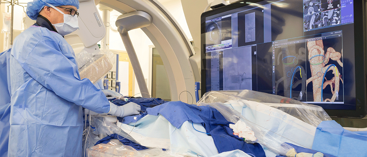
A novel imaging device recently rolled out at UT Southwestern is making complex aortic repairs safer for patients and operating room staff by dramatically reducing their exposure to radiation.
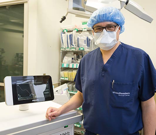
The device, known as Fiber Optic RealShape (FORS) and manufactured by Philips, uses light to visualize blood vessels, nearly eliminating the need for X-rays typically used during minimally invasive vascular procedures.
“Complex aortic repairs tend to be long operations that require frequent in-procedure imaging. Every time a surgeon presses on the X-ray pedal, the patient and staff – including assistants, nurses, scrub techs, anesthetists, and X-ray techs – get a dose of radiation,” said Carlos Timaran, M.D., Professor of Surgery and Chief of Endovascular Surgery. “The safety of each of these individuals is paramount, so reducing radiation exposure during these procedures is an important goal.”
UT Southwestern was one of less than a dozen medical centers in the U.S. and Europe chosen to participate in the early implementation of FORS due to its expertise and high volume of complex aortic repairs, said Dr. Timaran, who specializes in this procedure. He recently completed his 300th fenestrated endovascular aortic repair as part of his physician-sponsored investigational device exemption study, in which a patient-specific graft is used to support the aorta and its main branches.
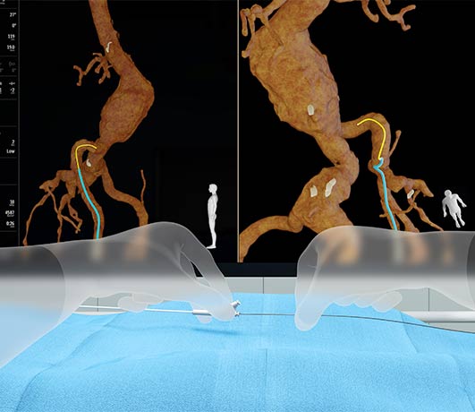
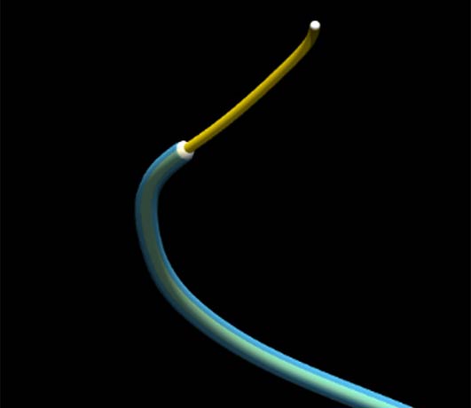
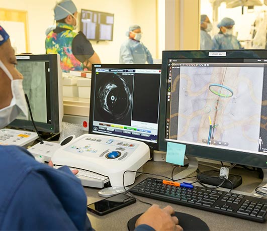
As an alternative to conventional imaging, the FORS device uses light traveling through hair-thin optical fibers built in specially designed catheters and wires to display its position and shape inside the body. Once this device is placed within a blood vessel, the strain on the optical fibers changes the light’s pathway. By analyzing how light reflects along the fibers, a computer algorithm reconstructs and visualizes the full shape of the device. The result is a real-time, three-dimensional view of the blood vessel that surgeons can overlay on computed tomography images taken before the procedure, providing a roadmap that surgeons can view from any angle to guide the surgery. Dr. Timaran said that far fewer X-rays are necessary when using FORS, significantly reducing exposure to patients and staff.
He expects FORS use to expand to other vascular applications over time. “This technology could potentially be used for any cardiovascular procedure,” said Dr. Timaran, who is a consultant for Philips. “This will eventually be the goal.”
UT Southwestern is ranked No. 11 in the nation for cardiology and heart surgery by U.S. News & World Report and nationally rated for its expertise in abdominal aortic aneurysm repair.
Dr. Timaran holds the Sam. H. Phillips, Jr. M.D. Distinguished Chair in Surgery.