

7 Tesla MRI discoveries shed new light on brain and skeletal muscle function
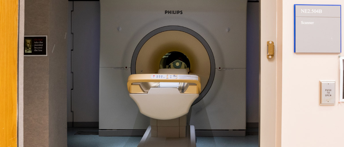
Only about 30 institutions in the U.S. have a 7 Tesla MRI, one of the most powerful imaging machines in existence with the ability to image less than a millimeter of tissue. The Advanced Imaging Research Center at UT Southwestern acquired one of these 24-ton giants of technology in 2007 to advance the Center’s work with magnetic MRIs and uphold the promise that higher quality imaging brings to science. The Center prioritizes this innovative imaging with harmless magnets and radio waves for its increasingly detailed images without the invasive surgery or possibly harmful radiation or X-rays. Jimin Ren, Ph.D., Associate Professor of Radiology and in the Advanced Imaging Research Center, and his colleagues used the machine to discover that:
Cellular energy metabolism is regulated by the balance of the oxidized and reduced forms of nicotinamide adenine dinucleotide (NADH and NAD+). Their content and ratio (NAD+/NADH, termed redox state), reflecting aging and oxidative stress, can now be measured in the human brain using the high-resolution 7T 31P MRS technique with spectral deconvolution method. The findings were published in the journal Magnetic Resonance in Medicine.
Cell membrane proteins and lipids are commonly decorated with a variety of sugar molecules to carry out crucial functions such as cell recognition and immune responses against viruses. Synthesis of these glycoproteins and glycolipids requires and is regulated by nucleotide sugars. Four such nucleotide sugars — uridine diphosphoglucose (UDP-glucose), UDP-galactose, UDP-N-acetyl-glucose, and UDP-N-acetyl-galactose — can be simultaneously measured in the human brain for the first time using the 31P MRS technique at 7T. The findings were published in the journal NMR and Biomedicine.
Normal energetic function of mitochondria relies on a well-regulated, demand-driven Krebs cycle. The metabolic rate of this cycle is regulated by the ratio of acetyl-CoA/free CoA through a coupled buffer system containing acetyl-carnitine and free carnitine. For the first time, the dynamic change of these two carnitine species can now be monitored in real time in human skeletal muscle during exercise using the high-resolution 1H MRS technique at 7T. The findings were published in NMR and Biomedicine.
Adequate ATP energy supply is essential for the body’s normal function, and a compromised energy production is implicated in a variety of human diseases including insulin resistance, Type 2 diabetes and Alzheimer’s disease. A novel method termed EBIT (exchange kinetics by band inversion transfer) had been developed on the human 7T scanner to allow noninvasive and simultaneous measurement of de novo ATP synthesis, creatine kinase activity, and cellular ATP motional dynamics in the human brain and skeletal muscle. The findings were published in NMR and Biomedicine.