

Maximum attraction
New Advanced Imaging Research Center Director drawn to latest upgrades for MRI facility
Dr. Anke Henning, the Director of a UT Southwestern research facility housing some of the world’s most sophisticated imaging equipment, rushes in for a meeting. On this day, she combines lunch with an interview, finishes in time to make it to back-to-back meetings, then finalizes plans for a Grand Canyon hiking trip with her family.
It’s been a busy year since she arrived from the Max Planck Institute for Biological Cybernetics in Germany to take over as Director of UT Southwestern’s Advanced Imaging Research Center (AIRC). In that time, she’s focused on defining and implementing a research strategy that emphasizes the development of advanced human and animal imaging technologies and translates these to use in UTSW’s clinical and basic science departments.
To accomplish this goal, Dr. Henning wants to recruit five or six faculty members with the expertise to realign the Center’s focus and is adding staff to her lab. She also plans major improvements to the Center’s infrastructure, including the upgrade of existing and installation of new human and animal imaging instrumentation as well as renovation of laboratory and office space.
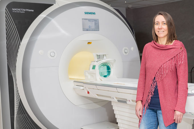
In her previous job in Tuebingen, Germany, Dr. Henning worked with one of the world’s three operational 9.4 tesla (9.4T) human whole-body MRI scanners, machines three times as strong as most patients encounter. In Dallas, she is applying for funding and negotiating with vendors to bring a 9.4T MRI to UT Southwestern, or maybe even one of the world’s first 11.7T super strength human MRI scanners.
Then there are the considerations to house such a powerful magnet. “It has to have 500 tons of steel shielding around it,” she says. It might go in next to the room holding the existing 7T, which is surrounded by 1-foot-thick steel walls, she says. (The AIRC installed the Southwest’s first 7T MRI machine for human research in 2007.)
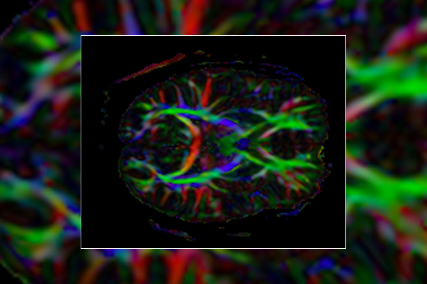
MRIs using such strong magnets – the magnet in a 7T is more than twice as powerful as those in clinical MRIs – can provide unprecedented detail in the resulting images.
The AIRC makes use of another strategy to enhance images. In the hyperpolarized MRI method, prior to a scan researchers inject polarized contrast agents that resemble naturally occurring metabolites, such as sugar.
The AIRC is also considering adding a small animal scanner to fully integrate positron emission tomography (PET) technology – which uses radioactive tracers to illuminate molecular activity inside the body – with an MRI to allow for PET/MRI readouts taken simultaneously. This addition would complement the Radiology Department’s soon-to-come installation of an integrated human PET/MRI scanner as well as its existing radiochemistry program.
Since its opening in 2005, AIRC researchers have worked to illuminate metabolic processes. They study how the body breaks down or builds needed substances when healthy and look at how those processes change in diseased cells, including in cancer cells that use far more energy than normal cells.
The anticipated development of ultra high field and hyperpolarized MRI, as well as integrated PET/MRI technology, will help continue this line of research. It will allow investigations of metabolic processes with high spatial and temporal resolution in live animals and humans rather than just metabolic readouts from isolated cells, tissues, or organs.
Training among giants
Dr. Henning, recruited to UT Southwestern as a Cancer Prevention and Research Institute of Texas (CPRIT) Scholar, sees the future Center as a place that designs and builds the latest technology for researchers. “I will focus on solving technologically challenging problems,” she says. “I would like to complement the Center with engineering that pushes the imaging research further. I’m really a physicist.”
She received her training at the Swiss Federal Institute of Technology (ETH) in Zurich, home to two researchers who won Nobel Prizes for their contributions to nuclear magnetic resonance (NMR), another technique that uses magnets to determine the chemical makeup of molecules.
Richard R. Ernst received a 1991 Nobel Prize in Chemistry for work that increased the sensitivity and resolution of NMR. Kurt Wüthrich shared a 2002 Nobel Prize in Chemistry for his role in the development of NMR spectroscopy, which lets researchers use magnetic fields to determine the structure of large biological molecules, such as proteins and fats, in solution. MRI uses that technology to create high-resolution anatomical and functional images of the animal and human brain and body. Magnetic resonance spectroscopy yields readouts showing the levels of various molecules in tissue.
Dr. Ernst had retired by the time Dr. Henning began her doctoral studies at ETH, although she attended seminars by him that she described as inspiring. When Dr. Wüthrich won his Nobel, she was there. “He was very happy when he won the Nobel Prize and ran across the campus giving a hug to anyone – including me,” she says. “I was in the department he headed and had classes under him. I learned my basic understanding of NMR spectroscopy there.”
After receiving her doctorate, Dr. Henning remained at ETH building her first research group. She joined the faculty of the Max Planck Institute for Biological Cybernetics in 2012.
While at ETH and Max Planck, Dr. Henning developed new MRI methods to “see” disease-related structural and metabolic changes in the brain, spinal cord, and heart.
Visionary ambitions
Her wish list at UTSW – upgrading to a 9.4T or 11.7T MRI – would allow researchers to look into nerve anatomy and brain function at the midrange, meaning the size of a molecule or cluster of atoms. That would support investigating metabolic adaptations faster and at higher spatial resolution. “We could image ions such as potassium, sodium, or calcium, and metabolites such as glutamate and GABA that are involved in neurotransmissions in the brain,” she says.
That might make it possible to see the underpinnings of psychiatric illnesses such as depression and neurodegenerative diseases like Parkinson’s and Alzheimer’s.
Basic neuroscience would benefit if the stronger MRI makes neurons and the long axon nerve fibers in the brain’s circuitry visible. “The question is,” she says, “can we push the resolution to an almost cellular level?” Another intriguing application might be the detection of currents that travel along nerve fibers. An 11.7T MRI might even make it possible to see changes taking place in the amino acids of a protein such as tau, which is implicated in Alzheimer’s, she says.
Another goal is translating the Center’s findings into new or improved treatments. For example, pinpointing metabolic abnormalities in a patient diagnosed with depression might let doctors predict which therapy would work best. And a closer look into neurodegeneration could show if a patient’s memory loss is the beginning of Alzheimer’s – or just normal aging.
“The question is, can we push the resolution to an almost cellular level?”
In other areas, high resolution imaging might permit precise neurostimulation of nonfunctioning neurons in patients with movement disorders to restore mobility. Or it might become possible to see the brain-heart interaction that leads to a malfunction such as broken heart syndrome, which mimics a heart attack, she says.
Putting AIRC tools to work
UT Southwestern’s 7T human MRI scanner was one of only a dozen or so in the world at the time it was installed, says Dr. Dean Sherry, founding Director of the AIRC and Professor of Radiology. Funding came from the Department of Defense.
Among the 7T’s users are Dr. Benjamin Greenberg, Professor of Neurology & Neurotherapeutics and Pediatrics as well as a Distinguished Teaching Professor, and Dr. Carol Tamminga, Professor and Chair of Psychiatry. Dr. Greenberg looks into treatments for Parkinson’s and multiple sclerosis, while Dr. Tamminga works to understand psychiatric disorders.
“Patients enrolled in our research studies take a new drug; we use an MRI technique that can identify the chemical molecules in a tissue and then we try to measure how the drug changes the functioning of the brain cells. It’s amazing technology,” Dr. Greenberg says.
Dr. Jimin Ren, Associate Professor of Radiology in the AIRC, developed the technology that lets Dr. Greenberg and others see such changes.
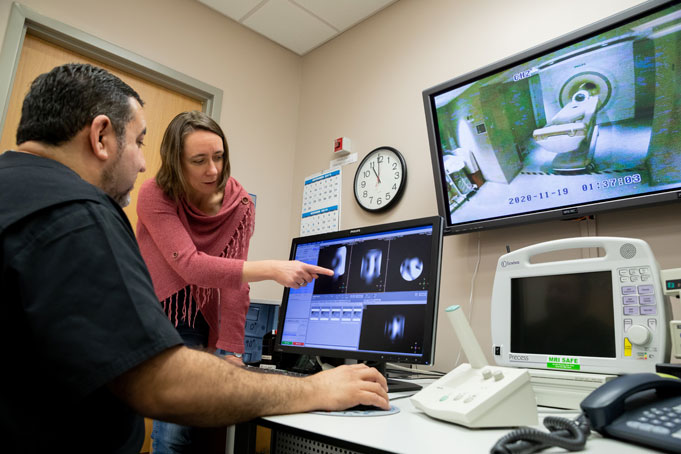
Over the years, Dr. Ren and his colleagues have scanned 2,200 research volunteers with the 7T. He positions volunteers onto the tan plastic conveyor belt-style bed, then pushes them into the large donut-shaped opening. Behind the “donut” is a superconducting electromagnet housed in a large steel cylinder that applies a magnetic field strength 140,000 times greater than that exerted by the Earth. A computer in the adjoining room receives the radio frequency signals emitted as magnetic moments of atomic nuclei in the volunteer’s body realign to the magnetic field after initial excitation by radio waves.
From European to Texan
As Dr. Henning juggles her plans for the Center, she is also adjusting to Texas life.
The woman who grew up behind Communism’s Iron Curtain until the fall of the Berlin Wall when she was 11 just got her first car – a Nissan. Biking was the fastest way to get around compact German and Swiss cities, she explains.
She marvels at the vast sparseness of the Lone Star State, a place with almost twice the land mass but less than a third the population of her native Germany.
Dr. Henning and her husband made it to the Texas State Fair last fall. “It was a bit of an odd mixture between animals, cars, and carousels,” she recalls. “My husband was fascinated, mainly by the car show.”
And the food? “We were amazed by some of the weird food, but we didn’t really try all of it. We avoided corn dogs and got frozen bananas.”
Dr. Greenberg is a Cain Denius Scholar in Mobility Disorders.
Dr. Sherry holds the Cecil H. and Ida Green Distinguished Chair in Systems Biology Science (UT Dallas).
Dr. Tamminga holds the Communities Foundation of Texas, Inc. Chair in Brain Science and the Lou and Ellen McGinley Distinguished Chair in Psychiatric Research.