

Radiology faculty artfully assist DMA to scan the unseen components of a Senufo helmet mask
Beauty, they say, is in the eye of the beholder. With the help of advanced imaging from UT Southwestern, however, the inner beauty and unseen intricacies of a Senufo helmet mask from the Dallas Museum of Art’s African collection are being appreciated more than ever before.
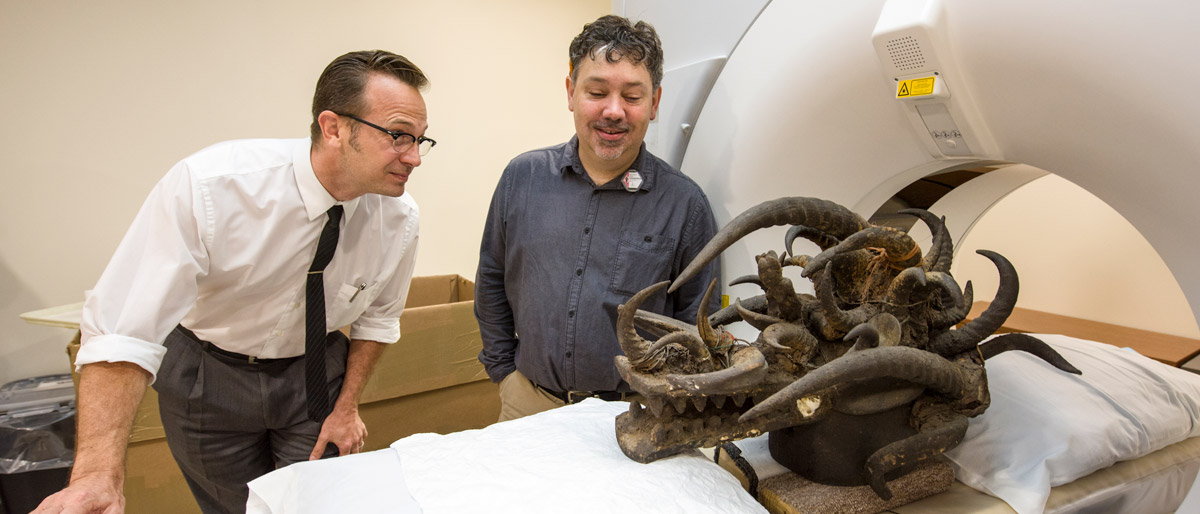
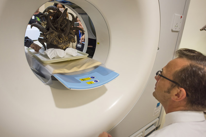
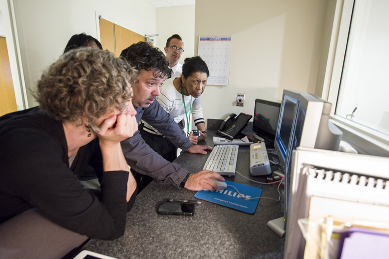
Dr. Matthew A. Lewis, Assistant Professor of Radiology, straddled the art-medicine-science border while imaging the helmet mask with a Philips IQon dual-layer detector spectral computed tomography scanner that was going through its pre-release campus testing. Clinically, an IQon is used 20 to 30 times daily for imaging of patients at William P. Clements Jr. University Hospital.
Related video: Inside a Senufo helmet mask
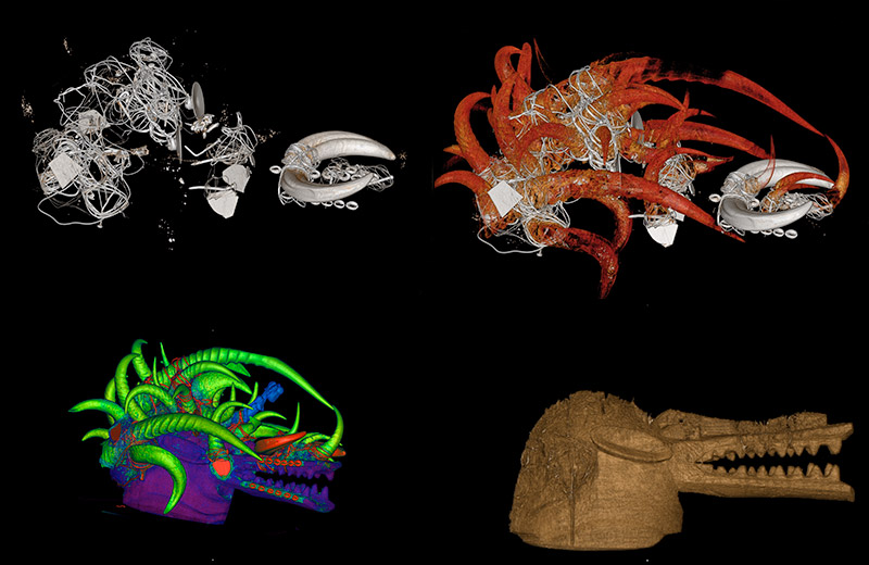
The resulting 3D images provided the foundation for the DMA’s “Not Visible to the Naked Eye: Inside a Senufo Helmet Mask” exhibit, which runs throughout this year. Images from this cutting-edge scanning technology provide museum visitors a deeper understanding of the mask-making process, its intricacies, and a detailed examination of materials and substances not visible to the naked eye. Those materials include buried animal skeletons, seeds, and metal underpinnings.
“Using the manufacturer’s IntelliSpace Portal as well as custom tools that we developed, we were able to better distinguish materials in the mask than would be possible with conventional CT,” said Dr. Lewis, who worked with colleagues at UTSW, UT Dallas, and the Dallas Zoo on the project.
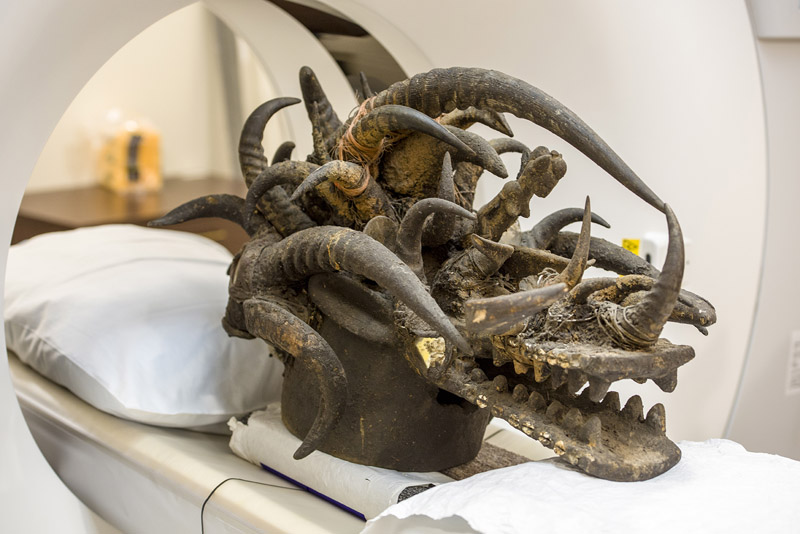
A Senufo helmet mask is worn by a medium at initiations, funerals, harvest celebrations, and secret events conducted by the powerful male-only Komo society, which has traditionally maintained social and spiritual harmony in Senufo villages in Côte d’Ivoire, Mali, and Burkina Faso in West Africa. The DMA’s mask – a 1997 donation to the collection from David T. Owsley – is ornately decorated with and empowered by 34 animal horns, a female figure, cowrie shells, and imported glassware. The CT scans further reveal unexpected materials and structures beneath the surface and objects contained in the horns.
Related photos: Radiologists capture the Senufo helmet mask
On March 18, museum curator Dr. Roslyn Adele Walker will discuss the community collaboration and the subsequent findings in a special 12:15 p.m. presentation in the DMA’s Conservation Gallery.
UT Southwestern medical students enrolled in “The Art of Examination” course first saw the mask on tours led by UTD’s Bonnie Pitman, a Distinguished Scholar in Residence and a national leader in the public engagement of art. Ms. Pitman’s work with The Edith O’Donnell Institute of Art History at UTD focuses on ways museums use their collections in developing close observation of works of art to enhance the diagnostic skills needed for medical practice.
Radiology’s involvement subsequently developed through discussions with UTD’s Dr. David McPhail, former Distinguished Chair of Conservation Science at the O’Donnell Institute and Professor of Chemistry.
“I wanted to tell him about the spectral CT scanner that Dr. Todd Soesbe and I were working with to see if there were any non-medical applications,” Dr. Lewis said. “He told me about the DMA wanting to image the mask. Since Dr. Soesbe and I were doing a lot of technical development on the clinical spectral CT at the time, we arranged for the imaging session and analyzed the imaging data.”
It’s an effort the UTSW team undoubtedly would do again.
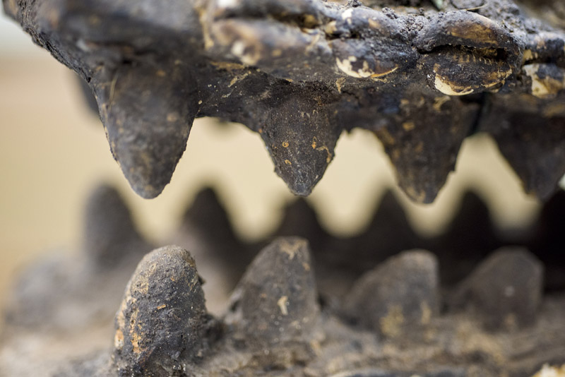
“It’s been really satisfying to apply our knowledge of imaging to help people in the art community who obviously appreciate and use science,” Dr. Lewis said. “I was especially happy when the tentative identification of the lizard skeleton inside the large lateral horn was confirmed, but many mysteries remain within the mask.
“What is the lump of metal in the bridge of the nose area? What are the other animals that are stuffed in the horns? What are the seed pods and other floral-looking items? How do we make the best artistic renderings of the mask that minimize the image artifacts due to all the metal nails and wire? It was all very exciting.”