

A head for brain science
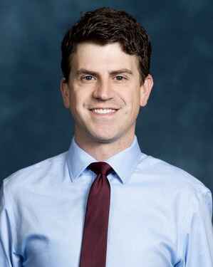
Research and compassion meet in Dr. William Dauer, Director of UT Southwestern’s O’Donnell Brain Institute.

Although the dirt is still flying on the new home for its nine-story, 300,000-square-foot research tower, the vision of the Peter O’Donnell Jr. Brain Institute’s inaugural Director, Dr. William Dauer, is starting to take shape.

Dr. Dauer, a physician-scientist, came to UTSW in July from the University of Michigan where he was Director of the Movement Disorders Group and of the Morris K. Udall Center of Excellence for Parkinson’s Disease Research. His research focuses on mechanisms of neurodegeneration in Parkinson’s disease and the molecular basis of the most common form of inherited primary dystonia in which patients experience involuntary twisting movements.
During his career, Dr. Dauer’s research has stretched from projects in his laboratory to multi-investigator team efforts like the one at the University of Michigan in which he still participates. That work involves searching for a therapy to prevent falls that severely impact the lives of people with advanced Parkinson’s.
One feature of Parkinson’s is the loss of the neurotransmitter dopamine in the striatum, an area of the brain responsible for helping us move through the world effortlessly, almost without thinking.
“Frequently, when I’m treating Parkinson’s patients, they will tell me without any prompting, ‘I have to just think about every movement, every step.’ The lack of dopamine makes moving effortful, requiring focused attention. However, dopamine loss occurs relatively early in the course of the disease, and patients who fall and those who don’t have similar degrees of dopamine loss,” says Dr. Dauer.
The falling problem arises from degeneration of a different set of nerve cells called cholinergic neurons, which are important for focusing attention. The lack of fluid movements from dopamine loss and reduced attention to guide movements tend to compound.
Researchers using animal models of Parkinson’s are working with clinicians and physician-scientists performing advanced brain imaging in patients to better understand how these factors combine to cause falls.
A class of drugs that stimulates the cholinergic cells has been approved to treat other conditions, and the hope is a member of that class may someday be used to prevent falls in people with Parkinson’s. Dr. Dauer stresses that clinicians carefully documenting the course of their patients’ illnesses led to the basic research underlying this work.
He hopes to inspire similar interdepartmental collaborations across clinical and basic science departments at UT Southwestern, where he has found many colleagues who share his enthusiasm for brain science.
Deep brain fascination
As long as he can remember, Dr. Dauer has been fascinated by the brain. As a boy in Southern California, he spent many happy hours playing with a plastic model of the brain. His grandfather was a pharmacist, and his father, uncle, and sister all became physicians.
“Being a physician was in the air in my house, and I just thought the brain was the coolest thing,” he recalls.
The effect of his early observations deepened his desire to treat all of his patients as individuals with a disease – not as diseased individuals.
Dr. Dauer learned firsthand about the power of the brain to impact lives. While he was growing up, his savvy, impressively well-educated mother developed multiple sclerosis. He noticed that people sometimes reacted to her physical disability by treating her as if she were mentally incapacitated.
The experience didn’t change his career trajectory; he was already sure about pursuing medicine. However, the effect of his early observations deepened his desire to treat all of his patients as individuals with a disease – not as diseased individuals. For him, that meant listening closely as patients described their day-to-day experiences in conversations that later informed his research. Those talks helped him understand the natural history of their illnesses.
Most of all, they influenced him as a teacher, inspiring him to convey the need to treat all patients with dignity, even if a person’s disease causes off-putting, involuntary twisting movements or tremors, as dystonias do. “The experience gave me the emotional energy to say, ‘Hey, this isn’t some flailing body, it’s a person.’ You have to develop a rapport with them,” he explains.
Dr. Dauer is known for his compassion as a clinician and for his dedication to teaching and learning. He envisions the O’Donnell Brain Institute as a place where medicine and science come together to advance discovery and patient care.
“Both kinds of insight are crucial,” he says.
Peeling the onion
An example of the need for layers of insight began in his independent research laboratory at Columbia University. After earning his M.D. from Washington University in St. Louis and completing an internship at Beth Israel Hospital in Boston, he became a neurology resident and fellow in movement disorders at Columbia.
While he was a fellow, others in the field identified the mutations underlying genetic forms of primary dystonia and Parkinson’s disease, prompting the young neurologist to pursue postdoctoral work in the Columbia laboratory of Dr. René Hen. There, Dr. Dauer learned how to genetically modify mice, creating some of the first knockout mice lacking the alpha-synuclein gene. He found that these mice were resistant to a toxin that can provoke Parkinson’s in humans, indicating that environmental and genetic forms of parkinsonism might act through common molecular pathways.
Beginning in 2001, Dr. Dauer published a series of papers establishing a function for the DYT1 dystonia protein torsinA at the nuclear membrane.
In following up these initial observations, the researchers created two more specific mouse models, one a knockout mouse that lacked the gene to make torsinA protein, the other a so-called knock-in mouse that had the human mutation in the corresponding mouse gene.
The similarity of the two lines of mice established that the DYT1 mutation impaired torsinA function. Both lines of mice died at birth, yet their nervous systems seemed fine. When the researchers studied the nerve cells’ nuclear membranes, however, they saw disordered areas that contained what looked like strange bubbles, known scientifically as blebs.
“We looked at every tissue we could and the abnormalities were only in nerve cells and confined in the nuclear membranes,” he says. “Here’s a protein that’s expressed in every cell in the body. It causes a neurologic disease, and when you either take it away or have two copies of the disease-causing mutation, you get a neural-selective, nuclear-membrane problem. That was really exciting because it said, ‘Hey, this protein has a special role in nerve cells.’”
“Why do blebs form in the nerve cells but not in the heart, muscle, or skin? That was the question that drove us at that time,” he recalls.
Those findings made them wonder about other members of the torsin family. Little was known about the five different forms of torsin that Dr. Dauer’s lab was studying except that levels of the various forms varied in different cell types.
“We asked: Could some of these other torsin family members compensate for missing torsinA in other cell types? If that was the case, then knocking down those other torsins would result in blebs like the ones we were seeing in nerve cells,” he explains.
The scientists knocked out different torsin family members, one by one, with striking results. Deleting torsinB – the family member with a sequence closest to that of torsinA and the one that naturally occurred at very low levels in nerve cells – did cause blebbing in nonneuronal cells.
Digging into torsin effects
The researchers’ next question: Could adding torsinB prevent the problem? Although they still lacked a mouse model, experiments in cultured cells indicated torsinB could prevent blebbing.
More interesting findings were on the way, and they depended heavily on matching clinical observations to laboratory developments.
In humans, DYT1 dystonia has a unique natural history. “The movement abnormalities in patients with DYT1 mutations only appear during later development – starting in late childhood or early adolescence. The symptoms worsen during a window of time spanning several years, and then remain static for life. These symptoms aren’t relentlessly progressive like Alzheimer’s or Parkinson’s, which indicates that torsinA plays a special role in the maturing brain, providing a clue for the development of mouse models of the disease that display the characteristic abnormal movements,” he says.
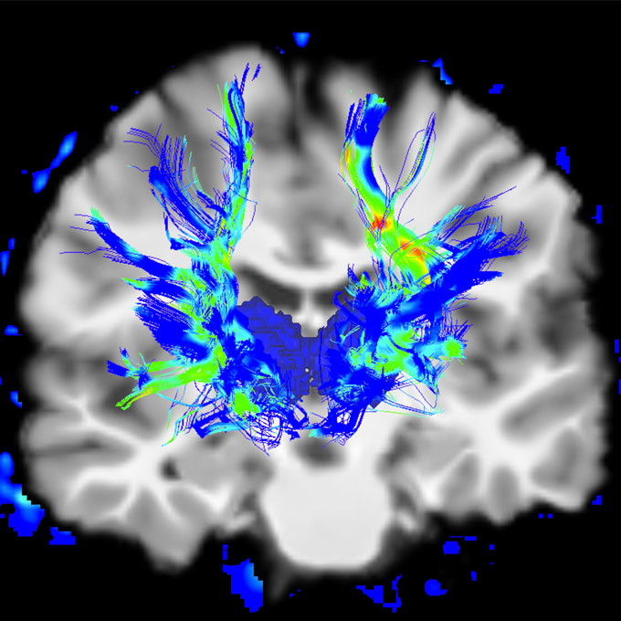
Credit: University of Michigan
After Dr. Dauer moved his lab to Michigan, a postdoc, Dr. Sam Pappas, informed by the natural history of the human disease, deleted torsinA from key areas of the brain in mice. In contrast to animals with complete loss throughout the brain, those with selective torsinA deletion survived. They also showed the pattern seen clinically in humans: seeming fine at birth, acquiring movement abnormalities as juveniles, and responding to therapies used clinically to treat humans.
Dr. Pappas, who recently joined UT Southwestern as an Assistant Professor and Associate Director of the Dauer lab, was first author of the eLife study showing that forebrain deletion of torsinA causes dystonic-like movements in mice.
“Sam is a terrific researcher with a gift for training graduate students and undergraduates. We always had five or six undergrads in the lab at Michigan,” Dr. Dauer says. “I hope to have some in my lab here, too.”
Another Michigan transplant is Jay Li, an M.D./Ph.D. student. “He’s doing some wonderful translational work,” Dr. Dauer says.
“He took the mouse model and asked, ‘What happens if we increase the levels of torsinB in the nervous system of this mouse? Will it affect movement? Will it do nothing? Will it be toxic?’” Dr. Dauer recalls, clearly thrilled to watch his student’s developing passion for research.
Work recently posted on BioRxiv reported dramatic findings: Overexpression of torsinB completely prevents abnormal movements. These findings point to a novel torsinB-based therapeutic strategy.
The future of brain science
Dr. Dauer envisions UT Southwestern researchers developing cross-departmental collaborations to ask and solve big, unanswered questions in neurodegenerative diseases, in brain repair mechanisms, and in mental illness.
In neurodegeneration and repair, investigations will stretch from the molecular level to the brain circuit and architecture level. “In many cases, we’re learning now, it’s not just what’s happening in the neurons,” he says. “It is increasingly clear that surrounding cells, like glial cells and immune system cells, all play a role in brain dysfunction.” Deciphering this puzzle will require scientists from diverse fields sharing insights to better understand the root causes of disease and how to prevent it.
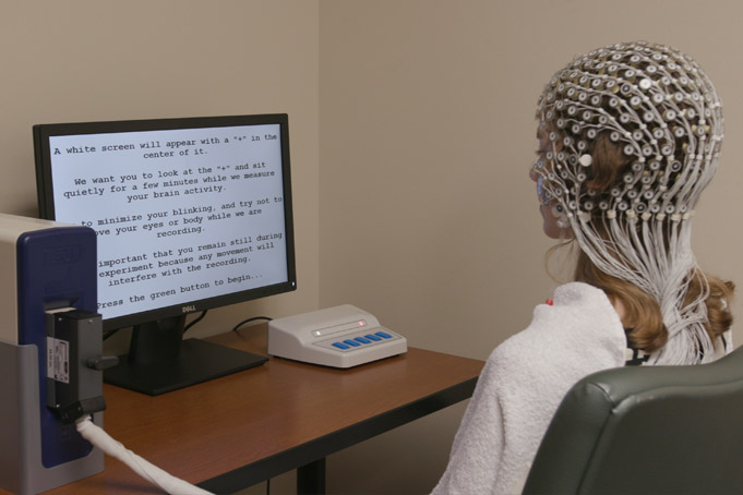
In psychiatric and neurologic disorders, experts are increasingly interested in reducing or preventing symptoms by modulating brain function. The most notable example is deep brain stimulation for Parkinson’s, in which small wires are implanted into the brain. But transcranial magnetic stimulation, high-intensity focused ultrasound (HIFU), and vagal nerve stimulation are other promising approaches in different diseases. The hope is that these or related experimental technologies may someday be used to treat disorganized brain function, movements, or behavior.
Researchers are just learning the very basics of how to modulate brain function from the outside, he says, whether with wires, sound waves, or magnetic pulses. Advances will require contributions from multiple disciplines, including individuals from clinical, basic science, engineering, and physics backgrounds.
“Through understanding the basic processes in the brain, we can identify chinks in the armor of these diseases and stop them,” he says. “That’s the hope.”
Dr. Dauer holds the Lois C.A. and Darwin E. Smith Distinguished Chair in Neurological Mobility Research.