


New 3-D fluorescence microscopes: Built from scratch, made for speed

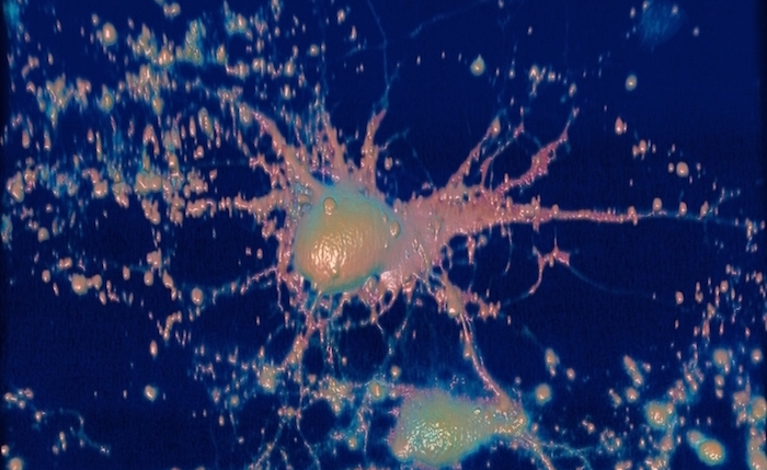
Bigger, faster, stronger. UT Southwestern researchers are developing microscopes powerful enough to see the future of medicine, today.
The study of living cells in three dimensions has been critically limited by the slow acquisition speed of current 3-D fluorescence microscopes, which assemble 3-D images from hundreds of two-dimensional images taken in a time-consuming serial process: One. After. Another. That approach is too slow to capture fast biological processes such as vesicular transport or the firing of neurons, says Dr. Reto Fiolka, an Assistant Professor in the Department of Cell Biology and in the Lyda Hill Department of Bioinformatics.
“To more closely mimic real-world conditions, we have abandoned traditional cell culture on hard, glass surfaces and moved toward 3-D environments.”
Dr. Fiolka led a UT Southwestern team that invented and built a 3-D fluorescence microscope for parallelized light-sheet fluorescence microscopy (pLSFM) that demonstrated a three-fold increase in image acquisition speed over previous light-sheet microscopes. In fact, the 3-D parallelized imaging scheme achieved, for every image plane, a performance comparable to a conventional single-plane microscope, the researchers reported in a study in the journal Optica in 2017.
“We realized that a conventional serial 3-D microscope runs into a bottleneck when trying to reach the fast rates of image acquisition necessary to observe some biological processes,” he says.
Low light is also a factor. Because cellular processes in the brain or deep in the body occur in darkness, the addition of light during imaging could damage the cells being imaged. Ideally, a faster imaging approach should not increase sample irradiance, he says.
The parallel approach developed in his laboratory overcomes the acquisition speed bottleneck, says Dr. Fiolka, a Cancer Prevention and Research Institute of Texas (CPRIT) Scholar. The parallelized system enabled researchers to accelerate the acquisition of the nearly 200 images in two dimensions needed to create clear 3-D images. The new system did all that without demanding a faster image detector and without needing brighter samples or more laser light than a conventional serial 3-D microscope would require, he notes.
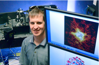
“We get three images at a time in parallel without losing light, meaning we can get the images three times as fast as would a serial microscope,” he says.
Dr. Fiolka’s lab continues to fine-tune the powerful microscope. In August, he and Dr. Kevin Dean, a postdoctoral researcher in Bioinformatics, published a refined approach for parallelized acquisition over larger volumes in Scientific Reports. The study was supported by grants from CPRIT and the National Institutes of Health.
“We are currently trying to simplify its design,” Dr. Fiolka says. “Simplification would help the dissemination of this work.”
Dr. Fiolka’s interest in imaging systems and lasers was sparked while he was an undergraduate in mechanical engineering during lectures in fluid dynamics and imaging techniques to measure the 3-D flow in a wind or water channel. “I was fascinated that one could image entire volumes at once with high precision. However, I saw more potential in the area of life sciences and biological research to develop new imaging technologies and switched my career path.”
Dr. Fiolka, who came to UT Southwestern following postdoctoral research at the Howard Hughes Medical Institute’s Janelia Research Campus, says the long-term goal of his laboratory is to develop and implement imaging technologies that provide unprecedented insight into cancer biology under conditions that are as close to the real world as possible.
“To more closely mimic real-world conditions, we have abandoned traditional cell culture on hard, glass surfaces and moved toward 3-D environments,” he says. “This requires continuous development of dedicated 3-D microscope technologies that minimize specimen photodamage and maximize optical penetration depth, image acquisition speed, sensitivity, and spatial resolution. We have made initial progress on all these fronts.”
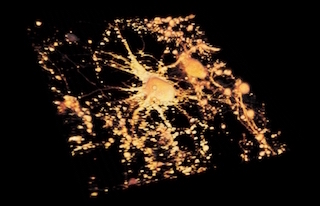
Since arriving at UT Southwestern, Dr. Fiolka has led or worked on teams that have developed six new 3-D imaging systems – each one designed to overcome specific problems in imaging, scale, or resolution encountered with current 3-D imaging technology, he says. Among his recent creations is a large-scale, long-term, light-sheet microscope that images cancer cell dissemination over whole zebrafish embryos or immune response in small organoids. Built to analyze large numbers of samples, it has enabled collaborations with the labs of Drs. James Amatruda, Rolf Brekken, and John Minna, he says.
“It is a noteworthy success story. I attribute some of it to its simplicity,” Dr. Fiolka says. “This new system can be used by collaborators with minimal training.”
Dr. Amatruda, Associate Professor of Pediatrics, Molecular Biology, and Internal Medicine, holds the Nearburg Family Professorship in Pediatric Oncology Research and is a Horchow Family Scholar in Pediatrics.
Dr. Brekken is a Professor of Surgery, Pharmacology, and in the Hamon Center for Therapeutic Oncology Research and an Effie Marie Cain Research Scholar.
Dr. Minna, Director of the Hamon Center for Therapeutic Oncology Research and Professor of Pharmacology and Internal Medicine, holds the Sarah M. and Charles E. Seay Distinguished Chair in Cancer Research and the Max L. Thomas Distinguished Chair in Molecular Pulmonary Oncology.