Low-oxygen environment leads to heart regeneration in mice, UTSW research shows
Update: Video added January 31, 2017.
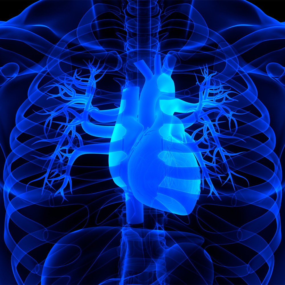
DALLAS – October 31, 2016 – Normal, healthy heart muscle is well-supplied with oxygen-rich blood. But UT Southwestern Medical Center cardiologists have been able to regenerate heart muscle by placing mice in an extremely low-oxygen environment.
Researchers with the Hamon Center for Regenerative Science and Medicine gradually lowered the oxygen in the air breathed by mice until it was at 7 percent – about the concentration of oxygen at the top of Mt. Everest. After two weeks in the low-oxygen environment, the heart muscle cells – called cardiomyocytes – were dividing and growing. Under normal circumstances cardiomyocytes do not divide in adult mammals.
The findings, published in Nature, build upon years of work that began with the discovery that the hearts of newborn mammals have the ability to regenerate, similar to the way skin has the ability to repair itself after a cut. But this ability of heart muscle to regenerate is quickly lost in the following weeks as the animal ages and cardiomyocytes are bathed in the oxygen-rich environment of the beating heart, causing damage to the cells.
“The adult human heart is not capable of any meaningful repair following a heart attack, which is why heart attacks have such a devastating impact,” said Dr. Hesham Sadek, Associate Professor of Internal Medicine and with the Hamon Center. “Though counterintuitive, we’ve shown that severely lowering oxygen exposure can sidestep damage to cells caused by oxygen and turn cell division back on, leading to heart regrowth.”
 Drs. Diana Canseco, Hesham Sadek, Wataru Kimura, and Yuji Nakada, part of the Hamon Center for Regenerative Science and Medicine, demonstrate a low-oxygen chamber, which was used to regenerate heart muscle in mice.")
In the current study, researchers lowered the oxygen level from the normal 21 percent to 7 percent over a period of weeks, then monitored the mass and function of the heart. They demonstrated that reduction in oxygen leads to both an increase in cardiomyocytes and improved heart function.
The researchers had tried a 10 percent oxygen environment, but there was no heart regrowth in the 10 percent oxygen environment. To avoid oxygen damage to cells, oxygen levels needed to be very low, a situation referred to as hypoxia.
“This work shows that hypoxia equivalent to the summit of Mt. Everest can actually reverse heart disease, and that is extraordinary,” said Dr. Benjamin Levine, Professor of Internal Medicine who holds the Distinguished Professorship in Exercise Sciences, and who directs the Institute of Exercise and Environmental Medicine at Texas Health Presbyterian Hospital Dallas, a joint program of UT Southwestern and Texas Health Resources.
“In theory, creating a low-oxygen environment could lead to repair not only of heart muscle, but of other organs as well,” said Dr. Sadek, who holds the J. Fred Schoellkopf, Jr. Chair in Cardiology. “Although exposure to this level of hypoxia can result in complications, it is tolerated in humans when performed in a controlled setting.”
The latest findings build upon previous research by UT Southwestern scientists that includes:
- A 2011 study in Science showing the ability of the neonatal mouse heart to regenerate
- A 2014 study in Cell in 2014 showing that oxygen metabolism causes damage to DNA in heart cells, which shuts down the ability to regenerate
- A 2015 study in Nature showing that a few heart cells which are in very low-oxygen pockets retain the ability to divide.
This work was supported by the National Institutes of Health, in addition to support from the Hamon Center for Regenerative Science and Medicine, whose goal is to understand the basic mechanisms for tissue and organ formation, and then to use that knowledge to regenerate, repair and replace tissues damaged by aging and injury. UT Southwestern established the Hamon Center for Regenerative Science and Medicine in 2014 with a $10 million endowment gift from the Hamon Charitable Foundation to further research into the relatively new field of regenerative medicine.
Other UT Southwestern researchers who contributed to the study are Dr. Yuji Nakada, Assistant Instructor of Internal Medicine; Dr. Diana Canseco, Instructor of Internal Medicine; SuWanee Thet, Research Associate; Dr. Salim Abdisalaam, postdoctoral researcher; Dr. Asaithamby Aroumougame, Assistant Professor of Radiation Oncology; and Dr. Wataru Kimura, Visiting Assistant Professor of Internal Medicine.
About UT Southwestern Medical Center
UT Southwestern, one of the premier academic medical centers in the nation, integrates pioneering biomedical research with exceptional clinical care and education. The institution’s faculty includes many distinguished members, including six who have been awarded Nobel Prizes since 1985. The faculty of almost 2,800 is responsible for groundbreaking medical advances and is committed to translating science-driven research quickly to new clinical treatments. UT Southwestern physicians provide medical care in about 80 specialties to more than 100,000 hospitalized patients and oversee approximately 2.2 million outpatient visits a year.
###
Media Contact: Cathy Frisinger
214-648-3404
cathy.frisinger@utsouthwestern.edu
To automatically receive news releases from UT Southwestern via email, subscribe at www.utsouthwestern.edu/receivenews