
2014 TIBIR Pilot Grant Recipients
Aberrant neurogenesis in a model of post-traumatic epilepsy
PI: Jenny Hsieh, Ph.D.

Department: Molecular Biology
Challenge
It is believed that a population of healthy neurons is generated after traumatic brain injury (TBI), possibly as a repair mechanism, in the hippocampus (a region responsible for learning and memory in the brain). However, there is also the formation of abnormal neurons called “aberrant neurogenesis” that may contribute to the disruption of normal circuits and post-traumatic epilepsy. We are trying to understand how aberrant neurogenesis after TBI promotes the recurrence of seizures and memory problems, which are long-term side effects in some patients.
Approach
To study the relationship between aberrant neurogenesis and seizures/memory, the Hsieh laboratory is developing an animal model of human post-traumatic epilepsy by using inbred mice induced with brain damage. Using their model, they will apply a genetic system to kill only the newly generated neurons or optogenetics using light to temporarily silence their activity. They will determine if epileptic mice with reduced aberrant neurogenesis have fewer recurrent seizures.
Overall Goals
To understand the process of new neuron formation after TBI, a process called “adult neurogenesis,” how it contributes to aberrant neurogenesis and the recurrence of seizures and associated memory problems.
Significance
This study could provide a basis for the treatment of post-traumatic epileptic seizures and seizure-related cognitive decline in patients by identifying aberrant neurogenesis as a clinically applicable target.
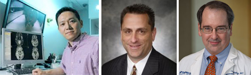
Neuronal regeneration in brain injury
 and his team of researchers studying the mechanisms of neuronal regeneration and recovery after brain injury. Front row from l to r: Tong Zang, Meng-Lu Liu; second row from l to r: right to left: Amel Ahmed, Yuhua Zou, Wenze Niu; top row from l to r: Dexiang Ban, Chao Guo.")
Department: Molecular Biology
Challenge
Injury to the brain leads to cell death, neuronal degeneration and the disruption of functional neuronal circuits. An unmet challenge in chronic brain injury is the replacement of lost neurons and restoration of normal brain function.
Approach
To address this challenge, the Zhang laboratory is taking a novel approach by reprogramming endogenous scar-forming, reactive glial cells into neural progenitors and functional neurons for brain regeneration and repair. Signaling pathways and epigenetic factors are being specifically examined for their role during the fate/neuronal reprogramming process
Overall Goals
To enhance the efficiency of in vivo reprogramming, promote maturation, survival and integration of newly generated neurons, and derive subtype-specific neurons for functional recovery of the adult central nervous system.
Significance
This study may give insight to the importance of neuronal replacement in functional recovery from brain injury.
Model of rotational acceleration-induced traumatic brain injury

PI: James Bibb, Ph.D.
Department: Psychiatry, Neurology and Neurotherapeutics
Challenge
Millions of sports- and war-related traumatic brain injuries (TBIs) occur annually in the U.S. These injuries are a result of the head undergoing rapid acceleration and deceleration, experiencing both linear and rotational forces. The brain is most sensitive to rotational motion, which causes shearing and a host of diffuse insults that can lead to long-term disability or even fatality. Unfortunately, there are limited preclinical models for rotation injuries after TBI and little insight to the mechanisms of rotational acceleration-induced TBI.
Approach
In a combined, interdisciplinary effort, Dr. Bibb’s lab is working with the UT Dallas Mechanical Engineering Department, and experts in the clinical diagnosis and treatment of TBI at UT Southwestern, including Jessica Moreland, M.D., in Pediatric Critical Care and Babu Welch, M.D., in Neurosurgery. Together, this team has developed a novel preclinical rodent model of rotational brain injury. Important guidance in this effort is also being provided by animal research experts at UT Southwestern, including Erin Jackson, D.V.M. (Veterinary Services), Stacy Pritt, D.V.M. (IACUC), and Shari Birnbaum, Ph.D. (Psychiatry Rodent Behavioral Facility).
Overall goals
To use the new model of rotational brain injury to assess the neuropsychiatric and neurological effects of these injuries and evaluate experimental treatments to protect from injury and counter their symptoms. In addition, Dr. Bibb wants to share their new TBI models with the research community and integrate their preclinical work with colleagues treating TBI patients and conducting clinical research.
Significance
This project could provide initial insights to the mechanisms of rotational acceleration-induced TBI and help identify treatments for TBI patients.
The predictive role of white matter injury for motor function recovery in a rat model of traumatic brain injury
PI: Hao Huang, Ph.D.
Department: Advanced Imaging Research Center, Radiology
Co-PIs: Robert Rennaker, Ph.D.; Craig Malloy, M.D.
Department: School of Behavioral and Brain Sciences, UT Dallas; Advanced Imaging Research Center, Internal Medicine, Radiology
Challenge
Moderate to severe TBI is known to impair balance and coordination and results in a loss of strength in humans, leading to prolonged motor dysfunction. To date no successful rehabilitative therapies have been developed and physical therapy results in incomplete recovery. Recovery of function following brain injury has been associated with cortical plasticity and reorganization. In order for the brain to reorganize connections, white matter (WM) must be intact and the degree of WM damage may be one factor that limits the ability of the brain to reorganize connections following TBI. A more complete understanding of the interaction between WM damage and functional recovery following TBI will serve to inform the development of rehabilitative strategies.
Approach
Previous studies demonstrated that vagal nerve stimulation (VNS) paired with rehabilitative training can significantly improve recovery of forelimb function compared to rehabilitative training alone in a rodent model of cortical ischemic stroke. Thus, Dr. Huang and his team will determine the relationship between WM injury and motor function recovery facilitated by VNS, measured by high resolution diffusion tensor imaging (DTI), hyperpolarized MRI and neuropathology in a controlled cortical impact rat model of TBI.
Overall Goals
To understand the WM pathology and predictive role of remaining WM in functional recovery from TBI and test the efficacy of VNS in TBI recovery.
Significance
Approved by FDA, delivery of VNS during rehabilitation is safe and has clear translational potential. Dr. Rennaker and his colleagues have completed a successful VNS clinical trial in Belgium and have begun an FDA-approved randomized, placebo-controlled study in Dallas. This study may reveal the changes in WM after TBI that can predict the efficacy of VNS therapies to enhance plasticity for recovery in TBI patients.
Bull Riders and Indirect Head Injury: A novel study aimed at a better understanding of rotational acceleration and indirect, whiplash-type shearing neurotrauma in sports-related brain injury
Department: Emergency Medicine
Challenge
Head injuries in athletes of all ages and skills, ranging from peewee sports to elite professional ballplayers, have recently been recognized as a significant and concerning health problem. These injuries range from concussion to severe TBI. Both linear and rotational accelerations are significantly correlated to concussion risk, but an acceptable animal or human model either in or outside the laboratory remains lacking.
Approach
Professional bull riders are elite athletes who experience some of the highest injury rates of any athlete in current day sports, with an alarmingly high rate of TBI. They experience rotational acceleration during the eight second bull ride via the whipping of the neck and the violent slinging of the head. This population allows the opportunity to evaluate this more subtle injury as a discrete event or multiple events over a short period of time. Dr. Wigginton will use sensor that measure linear and rotational response, measure biomarkers in the blood, short-term cognition and long-term neuropsychological performance.
Overall goals
To provide initial proof of concept that bull riders provide the cleanest and purest form of indirect, rotational acceleration head injury, thereby providing a previously unrealized opportunity to study this important component of head injury in sports.
Significance
This study could reveal a reliable model for further examination of the mechanisms of sports-related brain injury.