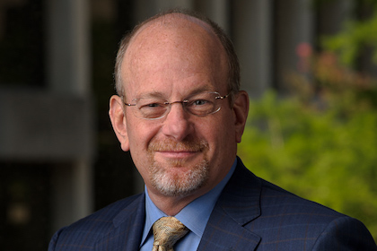
Biophysics Chair studies fundamental processes of T cell activation

By Deborah Wormser
UT Southwestern Medical Center Biophysics Chairman Dr. Michael Rosen spent part of his summer at the Marine Biology Laboratory at Woods Hole, Massachusetts, learning the steps in an intricate immune-system dance.
He collaborated with Dr. Ronald Vale, Professor of Cellular and Molecular Pharmacology at the University of California, San Francisco, and a recipient of the 2012 Lasker Prize. The result is a study recently published in the journal Science on fundamental processes in T cell activation. The two Howard Hughes Medical Institute (HHMI) Investigators are corresponding authors and the two lead authors, both postdoctoral researchers, are Dr. Xiaolei Su of UCSF and HHMI; and Dr. Jonathon Ditlev of the UT Southwestern Department of Biophysics and HHMI.
The human body is constantly assaulted by damaging agents – infectious bacteria, viruses, and even its own malignant cells in the case of cancer. The white blood cells, a central component of the immune system, maintain constant surveillance for these invaders, acting with great specificity to identify and attack them. T cells are a particular class of white blood cells that have several important roles in the immune system, Dr. Rosen explained.
For instance, when a T cell encounters a virally infected cell, the two cells stick to each other through interactions between proteins on the surface of each cell. These interactions then induce a complicated cascade of events inside the T cell, including stimulation of a series of proteins called kinases and rearrangement of a filament network called the actin cytoskeleton. These events ultimately cause the T cell to divide – creating new disease-fighting T cells – and also to secrete proteins that stimulate other elements of the immune system in a process called T cell activation.
“When scientists have observed T cell activation under a microscope, they have seen that many proteins inside the cell undergo a beautiful dance at the cell-cell interface, the place where the T cell and the virally infected cell stick together,” said Dr. Rosen. “At the very earliest stages, many of the proteins come together into small dots, roughly 1/20th the diameter of a human hair. These dots move around on the cell-cell interface and eventually coalesce into a large spot at the center of that interface.”
Although the proteins within the dots were long thought to be important for T cell activation, scientists have not understood exactly how the dots formed or how the dots contributed to T cell activation, he said.
Drs. Rosen and Vale worked to solve those mysteries, starting in the summer of 2013 as part of the HHMI’s interdisciplinary Summer Institute at Woods Hole. The research team decided to reconstitute the initial stages of T cell activation biochemically by purifying more than a dozen proteins or protein complexes known to assemble into dots at the cell-cell interface during T cell activation. They attached three of the proteins or complexes to an artificial T cell membrane, and the remainder were studied in solution.
When they mimicked T cell activation by adding a chemical energy source to the solution, all the proteins assembled into dots on the membrane, very similar to what is seen in T cells, Dr. Rosen said.
“Additional experiments showed that the proteins come together through a process called phase separation, which is similar to the separation of oil droplets from water when you shake salad dressing in a flask and watch the oil droplets clump back together. By comparing the behaviors of the proteins in our reconstituted system with those observed in T cells, we were able to show that the dots form in T cells through this same phase separation mechanism,” he said. “Further, we showed that the phase separation stimulates downstream events needed for T cell activation, including assembly of the actin cytoskeleton and activation of a key kinase,” he said.
Dr. Rosen explained that the work is significant for several reasons.
“First, it establishes how the protein dots form at the cell-cell interface during T cell activation. Given that many cellular processes are mediated by proteins similar to those in T cells, this mechanism is probably widespread in biology. Second, it reveals some of the biochemical and cellular functions of phase separation. Again, some of the functions we found for the dots produced during T cell activation may be general to other processes in other types of cells.
“Finally, to our knowledge, no one before has reconstituted such a complicated membrane-associated pathway from purified proteins. By doing so, we were able to address long-standing questions in the mechanism of T cell activation,” he said.
Dr. Rosen’s research partnership with Dr. Vale continues through the summer of 2017.
Other UT Southwestern authors were graduate student Wenmin Xing and Dr. Sudeep Banjade, a postdoctoral researcher, both of Biophysics. In addition, three more researchers from UCSF participated, as did one from the HHMI Mass Spectrometry Laborators and Department of Molecular and Cellular Biology at the University of California, Berkeley.