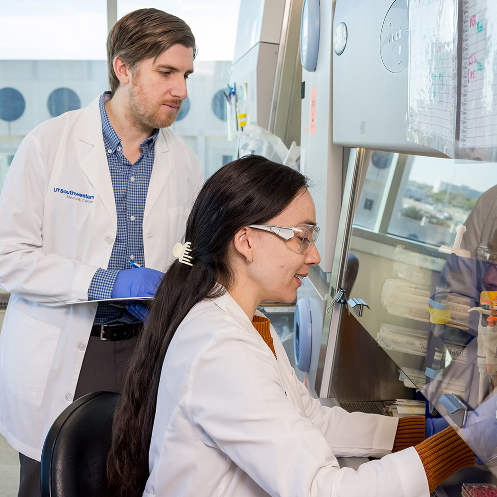
Advanced imaging driving CPRIT-supported projects

DALLAS – May 4, 2018 – Three grants of almost $4.2 million awarded to UT Southwestern faculty members will support image-based investigations into the metabolic pathways and basic processes of heart disease, prostate cancer, and breast cancer. In all, the Cancer Prevention and Research Institute of Texas (CPRIT) in late February awarded 49 research grants and eight prevention grants totaling more than $73.5 million across the state in this year’s initial round of support.
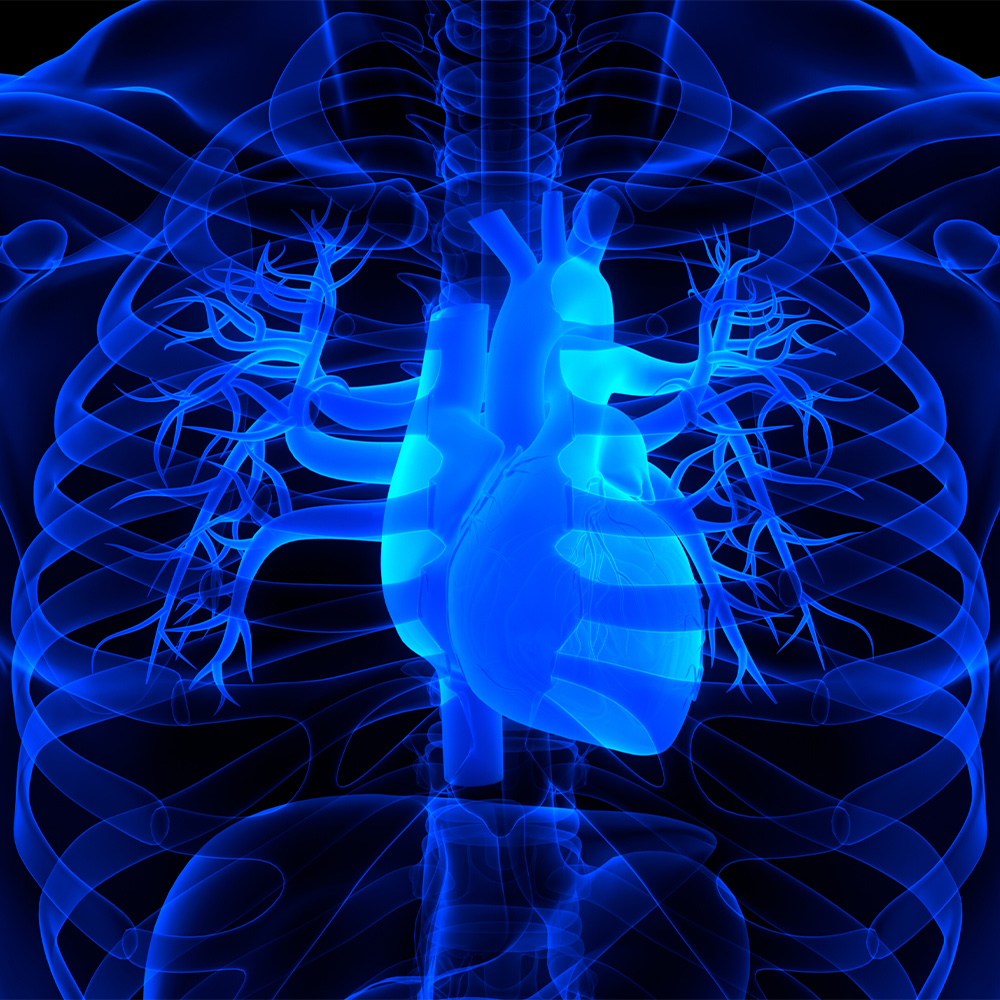
Dr. Vlad Zaha, Assistant Professor of Internal Medicine, received almost $2.4 million to delve into noninvasive detection of anthracycline-induced cardiotoxicity using hyperpolarized carbon-13-based magnetic resonance spectroscopic imaging. Dr. Zaha, one of the few onco-cardiologists in the Dallas area, provides cardiovascular care for cancer patients and cancer survivors.
The National Cancer Institute estimates that there will be about 18 million cancer survivors in the United States by 2022, many facing short- and long-term cardiovascular complications caused by today’s cancer chemotherapy or radiotherapy. In a recent National Health and Nutrition Examination Survey regarding the mortality of 1,807 cancer patients, 51 percent died of recurring cancers and another 33 percent of heart disease in the seven years following their diagnoses. Early detection and treatment of heart disease in cancer patients is critical to improve survival after cancer treatment.
“We are developing noninvasive, nonradioactive imaging modalities to identify the earliest biochemical changes that appear in heart disease,” Dr. Zaha said of his project at UT Southwestern’s Advanced Imaging Research Center (AIRC). “Our goal is to identify changes in the way the heart is transforming chemical energy before structural changes are setting in. Heart damage is a significant side effect of anthracyclines in patients who require this treatment. Anthracyclines start causing heart injury by damaging chemical energy transformation processes. We aim to measure these effects and develop a new, early diagnostic modality for heart disease.”
Utilizing the AIRC’s hyperpolarization magnetic resonance imaging (MRI) suite will allow Dr. Zaha’s team to track fundamental, discrete steps in the energetic metabolism of the heart. They say that overlaying this information in turn on a regular cardiac MRI will allow them to generate a dynamic, real-time map of the biochemical energy transfer processes in the heart.
“Carbon is the backbone of biochemical compounds such as carbohydrates, proteins, and fatty acids, which form the structure of the heart, but are also used in the heart for chemical energy transformations,” Dr. Zaha said. “The nuclei of carbon-13, a nonradioactive carbon isotope, behave like small magnets due to their odd atomic number. Their resonance with the magnetic field can be used to track their transition from one molecule to another in a chemical process. With hyperpolarization, these signals can be enhanced more than 10,000-fold, which allows us to identify the chemical transition processes much faster and on a smaller scale, and to transform these signals into dynamic images of the biochemical activity in the heart.”

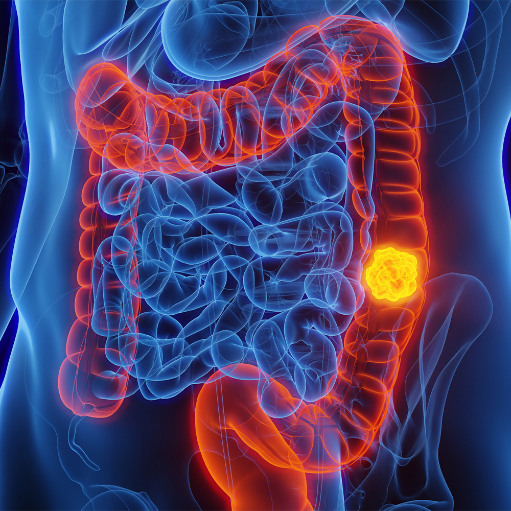
Drs. Dean Sherry and Elena Vinogradov each received $900,000 to pursue research related to early detection of prostate cancer and breast cancer, respectively.
Dr. Sherry, Director of the AIRC and a Professor of Radiology, will use MRI to detect glucose-stimulated zinc secretion from the prostate. The gland, he explained, stores large amounts of zinc ions when healthy, more than any other human tissue. When prostate cells become malignant, however, they lose their ability to accumulate zinc.
“In principle, if one could image zinc content, it would differentiate prostate cancer from a healthy prostate,” Dr. Sherry said. “Over the past few years, we developed a responsive MRI agent that brightens an image only when it encounters high levels of zinc. So the basis of our work is to translate this imaging technology from animal models to man.”
Multiparametric MRI is the usual method of first identifying prostate cancer in men whose PSA levels have risen above a medically accepted threshold. This imaging technique, Dr. Sherry said, works well in many cases, but the results can be confounding in certain zones of the gland where benign prostatic hyperplasia (BPH) and cancerous tissue appear similar by MRI.
“Ultimately, this leads to either an ultrasound or MRI-guided tissue biopsy, which is expensive and painful,” Dr. Sherry said. “Our hope is that this new agent might be able to differentiate prostate cancer from BPH tissues in a simple MRI exam and thereby allow one to monitor growth or shrinkage of prostate cancer with therapy.”
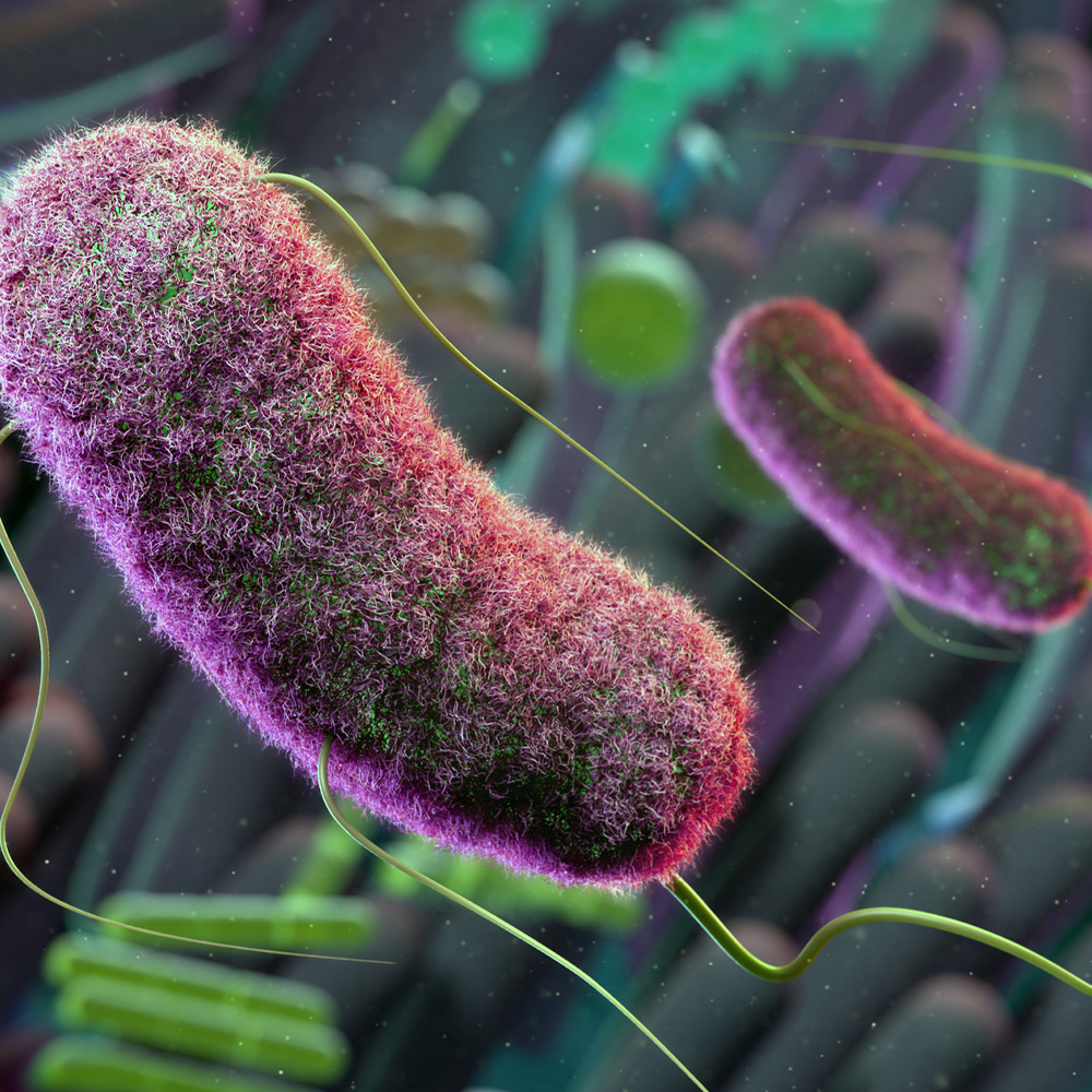
Dr. Vinogradov, Assistant Professor of Radiology, will use chemical exchange saturation transfer (CEST) MRI to detect biochemical alterations in human breast malignancy. Mammography is the current gold standard in breast cancer detection. But breast cancer management has recently come under scrutiny due to concerns about the increased number of unnecessary biopsies and unjustifiably aggressive treatments.
“We are developing a novel molecular CEST MRI method and tailoring it for breast malignancy assessment,” said Dr. Vinogradov. “CEST is sensitive at concentrations that are too low to impact the contrast of standard MRI.
“By merging advances in biochemistry with state-of-the-art imaging, our goal is to create a noninvasive and nonionizing imaging tool capable of detecting alterations in cancer biology at the biochemical and genetic level,” she added. “We hope that CEST will better differentiate malignant tumors from benign lesions, with further differentiation of aggressive tumors. For treatment planning, CEST MRI may noninvasively predict tumor therapy response, thus improving patient outcomes.”
To date, CPRIT has awarded 1,247 grants totaling more than $1.95 billion and is nearing 70 percent dispersal of the state’s historical 2007 constitutional commitment of $3 billion to fight cancer. During the 85th Texas Legislature, CPRIT’s Sunset Review date was extended by two years to 2023 to allow the agency to award all funds approved by Texas voters.
Dr. Sherry also is a Professor of Chemistry at UT Dallas, where he holds the Cecil H. and Ida Green Distinguished Chair in Systems Biology.
About UT Southwestern Medical Center
UT Southwestern, one of the premier academic medical centers in the nation, integrates pioneering biomedical research with exceptional clinical care and education. The institution’s faculty has received six Nobel Prizes, and includes 22 members of the National Academy of Sciences, 16 members of the National Academy of Medicine, and 14 Howard Hughes Medical Institute Investigators. The faculty of more than 2,700 is responsible for groundbreaking medical advances and is committed to translating science-driven research quickly to new clinical treatments. UT Southwestern physicians provide care in about 80 specialties to more than 100,000 hospitalized patients, 600,000 emergency room cases, and oversee approximately 2.2 million outpatient visits a year.