Illuminating cancer: Researchers invent a pH threshold sensor to improve cancer surgery
Update: Video May 1, 2017
DALLAS – December 20, 2016 – UT Southwestern Medical Center researchers have invented a transistor-like threshold sensor that can illuminate cancer tissue, helping surgeons more accurately distinguish cancerous from normal tissue.
In this latest study, researchers were able to demonstrate the ability of the nanosensor to illuminate tumor tissue in multiple mouse models. The study is published in Nature Biomedical Engineering.
“We synthesized an imaging probe that stays dark in normal tissues but switches on like a light bulb when it reaches solid tumors. The purpose is to allow surgeons to see tumors better during surgery,” said senior author Dr. Jinming Gao, Professor of Oncology, Pharmacology and Otolaryngology with the Harold C. Simmons Comprehensive Cancer Center.
The nanosensor amplifies pH signals in tumor cells to more accurately distinguish them from normal cells.
“Cancer is a very diverse set of diseases, but it does have some universal features. Tumors do not have the same pH as normal tissue. Tumors are acidic, and they secrete acids into the surrounding tissue. It’s a very consistent difference and was discovered in the 1920’s,” said Dr. Baran Sumer, Associate Professor of Otolaryngology, and co-senior author of the study.
The researchers hope the improved surgical technology can eventually benefit cancer patients in multiple ways.
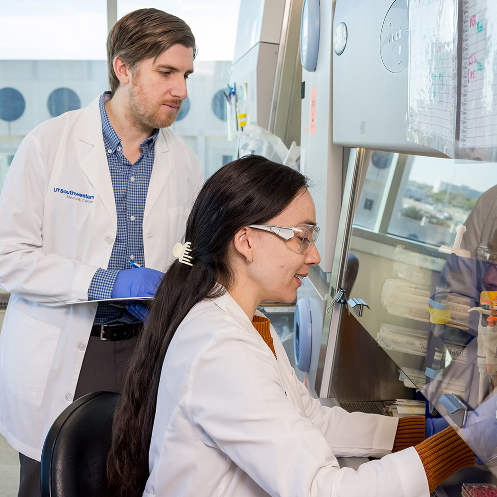
 and Dr. Jinming Gao (right) and have invented a transistor-like threshold sensor that can illuminate cancer tissue, helping surgeons more accurately distinguish cancerous from normal tissue in mouse models.")
“This new digital nanosensor-guided surgery potentially has several advantages for patients, including more accurate removal of tumors, and greater preservation of functional normal tissues,” said Dr. Sumer. “These advantages can improve both survival and quality of life.”
For example, this technology may help cancer patients who face side effects such as incontinence after rectal cancer surgery.
“The new technology also can potentially assist radiologists by helping them to reduce false rates in imaging, and assist cancer researchers with non-invasive monitoring of drug responses,” said Dr. Gao.
According to the National Cancer Institute, there are 15.5 million cancer survivors in the U.S., representing 4.8 percent of the population. The number of cancer survivors is projected to increase by 31 percent, to 20.3 million, by 2026.

Dr. Sumer and Dr. Gao were joined in this study by Dr. Gang Huang, Instructor of Pharmacology; Dr. Xian-Jin Xie, Professor of Clinical Sciences; Dr. Rolf Brekken, Professor of Surgery and Pharmacology and an Effie Marie Cain Research Scholar; and Dr. Xiankai Sun, Director of Cyclotron and Radiochemistry Program in Department of Radiology and Advanced Imaging Research Center, Associate Professor of Radiology, and holder of the Dr. Jack Krohmer Professorship in Radiation Physics; Dr. Joel Thibodeaux, Assistant Professor of Pathology and Director of Cytopathology, Parkland Memorial Hospital.
Additional UT Southwestern researchers who contributed to the study include: Dr. Tian Zhao, Dr. Xinpeng Ma, Mr. Yang Li, Dr. Zhiqiang Lin, Dr. Min Luo, Dr. Yiguang Wang, Mr. Shunchun Yang and Ms. Zhiqun Zeng in the Harold C. Simmons Comprehensive Cancer Center; and Dr. Saleh Ramezani in the Department of Radiology.
Dr. Gao and Dr. Sumer are scientific co-founders of OncoNano Medicine, Inc. The authors declare competing financial interests in the full-text of the Nature Biomedical Engineering article. UT Southwestern Medical Center has licensed the technology to OncoNano Medicine and has a financial interest in the research described in the article.
Funding for the project includes grants from the Cancer Prevention and Research Institute of Texas. Dr. Gao and Dr. Sumer are investigators for two Academic Research grants and OncoNano Medicine was the recipient of a CPRIT Product Development Research grant.
Research reported in this press release was supported by the National Cancer Institute under Award Number R01 CA192221 and the National Institute of Biomedical Imaging and Bioengineering of the National Institutes of Health. The content is solely the responsibility of the authors and does not necessarily represent the official views of the National Institutes of Health.
The Harold C. Simmons Comprehensive Cancer Center is the only NCI-designated Comprehensive Cancer Center in North Texas and one of just 47 NCI-designated Comprehensive Cancer Centers in the nation. Simmons Cancer Center includes 13 major cancer care programs. In addition, the Center’s education and training programs support and develop the next generation of cancer researchers and clinicians. Simmons Cancer Center is among only 30 U.S. cancer research centers to be designated by the NCI as a National Clinical Trials Network Lead Academic Participating Site.
About UT Southwestern Medical Center
UT Southwestern, one of the premier academic medical centers in the nation, integrates pioneering biomedical research with exceptional clinical care and education. The institution’s faculty includes many distinguished members, including six who have been awarded Nobel Prizes since 1985. The faculty of almost 2,800 is responsible for groundbreaking medical advances and is committed to translating science-driven research quickly to new clinical treatments. UT Southwestern physicians provide medical care in about 80 specialties to more than 100,000 hospitalized patients and oversee approximately 2.2 million outpatient visits a year.
###
Media Contact: Lori Sundeen Soderbergh
214-648-3404
lori.soderbergh@utsouthwestern.edu
To automatically receive news releases from UT Southwestern via email, subscribe at www.utsouthwestern.edu/receivenews