Radiology blazes new trails, as department celebrates 60th anniversary

As UT Southwestern’s Department of Radiology celebrates its 60th anniversary, the team continues to blaze new trails in imaging and radiological services, including the opening this past year of its first cyclotron facility that generates radioisotopes and tracers for biomedical research and cancer treatments.
Supported by the Cancer Prevention and Research Institute of Texas (CPRIT), the Cyclotron and Radiochemistry Program leverages Positron Emission Tomography (PET), which is especially suited for understanding cancer initiation and progression, cancer cell metabolism, prognostic evaluation of cancer patients, and eventually the early and individualized diagnosis of cancer.
Underground on UT Southwestern’s North Campus, the cyclotron facility generates positron-emitting radioisotopes and radiotracers that enable preclinical, translational, and clinical research with PET imaging. This research, which crosses many disciplines, accelerates the translation of basic research discoveries from bench to bedside for better patient care and positive clinical outcomes. The Cyclotron and Radiochemistry Program also serves as an educational resource to promote the integration of advanced imaging technologies into disease biology, prevention, and therapeutic monitoring.
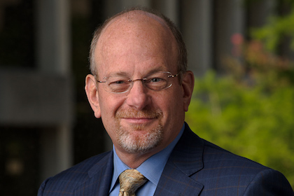
“The UT Southwestern Medical Center Department of Radiology has a proud legacy of excellence and innovation. We are currently in one the most exciting times in our Department’s history, with unprecedented expansion and opportunity that mirror the growth and opportunity afforded by the Dallas-Fort Worth area,” said Dr. Neil Rofsky, Professor and Chairman of Radiology, and Director of Translational Research for UT Southwestern’s Advanced Imaging Research Center.
The Department’s beginning traces back to Dr. Frederick Bonte, now Professor Emeritus, who was appointed the first Chairman of Radiology in 1956. Previous to that, UT Southwestern radiology services had been provided as part of the Department of Surgery.
Dr. Bonte, who later became Dean of UT Southwestern Medical School, set the bar high from the start through a distinguished career that has furthered the clinical use of various radiologic tools and techniques. Those techniques included single-photon emission computed tomography (SPECT) to identify a characteristic sign of Alzheimer’s disease and distinguish it from a group of illnesses known as frontotemporal diseases. Other work of Dr. Bonte included a study that was the first to pinpoint damage inside the brains of veterans suffering from Gulf War syndrome.
In 1977, Dr. Robert Parkey succeeded Dr. Bonte as Chair of Radiology. Along with Dr. Bonte, he was instrumental in developing a technique called technetium-99m stannous pyrophosphate imaging to detect acute myocardial infarction, used worldwide to diagnose, localize, and determine the size of acute heart attacks.
Today, breakthrough radiology research and advancement in imaging techniques is spread across nine clinical divisions: Abdominal Imaging, Breast Imaging, Cardiothoracic Imaging, Vascular Interventional Radiology, Magnetic Resonance Imaging, Musculoskeletal Imaging, Neuroradiology/Interventional, Nuclear Medicine, and Pediatric Radiology. Recent recruits of chiefs for seven of these divisions have strengthened the Department’s complement of clinician-scientists. Furthermore, a dedicated research section within Radiology focuses on developing and improving image acquisition and reconstruction techniques, enabling a better understanding and evaluation of a variety of disease processes.
Members of the Department of Radiology perform more than 900,000 exams annually across the Medical Center, including at William P. Clements Jr. University Hospital, Zale Lipshy University Hospital, Parkland Memorial Hospital, Children’s Medical Center Dallas, and at a handful of outpatient clinics, including the Outpatient Building, James W. Aston Ambulatory Care Center, Algur H. Meadows Diagnostic Imaging Center, Bill and Rita Clements Advanced Medical Imaging Building, and the Mary Nell and Ralph B. Rogers Magnetic Resonance Imaging Center.
The Clements Imaging Building is home to the Mary Nell and Ralph B. Rogers Magnetic Resonance Center, the Positron Emission Tomography (PET) Imaging Facility, and the Advanced Imaging Research Center.
The Rogers Magnetic Resonance Center is equipped with some of the latest technology available, including two state-of-the-art 3Tesla magnets that accommodate all types of scans and patients, including claustrophobic patients in one of the center’s wide-bore MRI scanners. Cutting-edge technology is applied to all types of scans, from routine MRI of joints to the most complex, neurologic, and cardiac studies. Advanced MRI capabilities are routinely applied for evaluation of patients with neurologic diseases and musculoskeletal disorders, as well as diseases of the chest, abdomen, and pelvis. MRI protocols also assist planning radiation treatments in the brain, spine, liver, kidney, prostate, and cervix.
The Meadows Imaging Center on South Campus serves both Parkland and UT Southwestern patients, offering advanced MRI techniques, such as spectroscopy and perfusion. It also includes an ultra-high-resolution 3T scanner.
The Positron Emission Tomography (PET) Imaging Facility features high-resolution PET scanning for neurological, oncological, and psychiatric applications. Unlike more routine imaging systems that show what tissues look like, such as X-ray, CT, ultrasound, or MRI, PET studies reveal information about tissue function and metabolism that are frequently used to diagnose and stage cancer.
The Advanced Imaging Research Center, known as AIRC, was created in 2005 through a collaboration between UT Southwestern and other North Texas institutions to further research in MRI techniques and translate those discoveries into clinical practice. The Center’s research seeks to advance basic understanding, diagnostic techniques, and treatments for a wide range of diseases, including cancer, diabetes, obesity, Alzheimer’s disease, schizophrenia, depression, autism, attention-deficit hyperactive disorder (ADHD), and diseases of the heart, lung, and liver. AIRC faculty also help train undergraduate and graduate students from UT Southwestern, UT Dallas, and UT Arlington, as well as international postdoctoral scientists.
###
Dr. Rofsky holds the Effie and Wofford Cain Distinguished Chair in Diagnostic Imaging.